Image Gallery
Confocal, STED super-resolution, and transmission EM images in one viewer. Filter by modality, swipe or use the arrow keys, and open any image full screen.
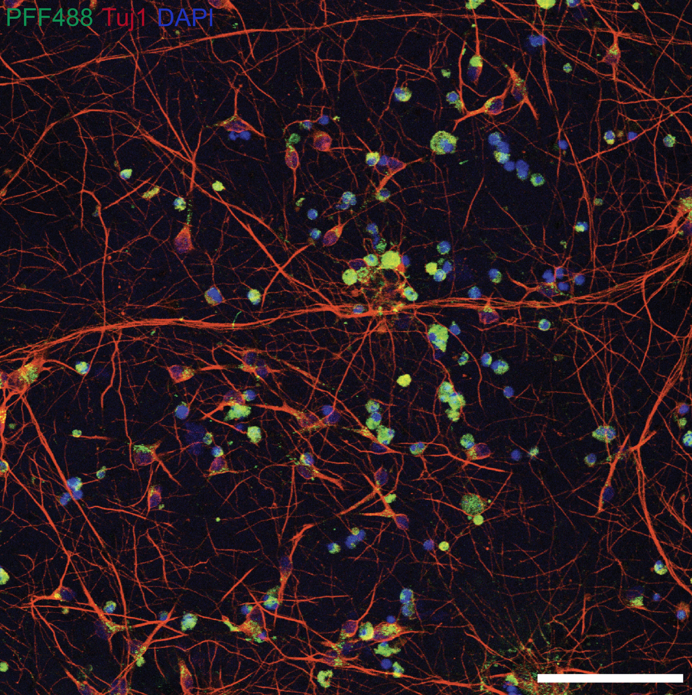
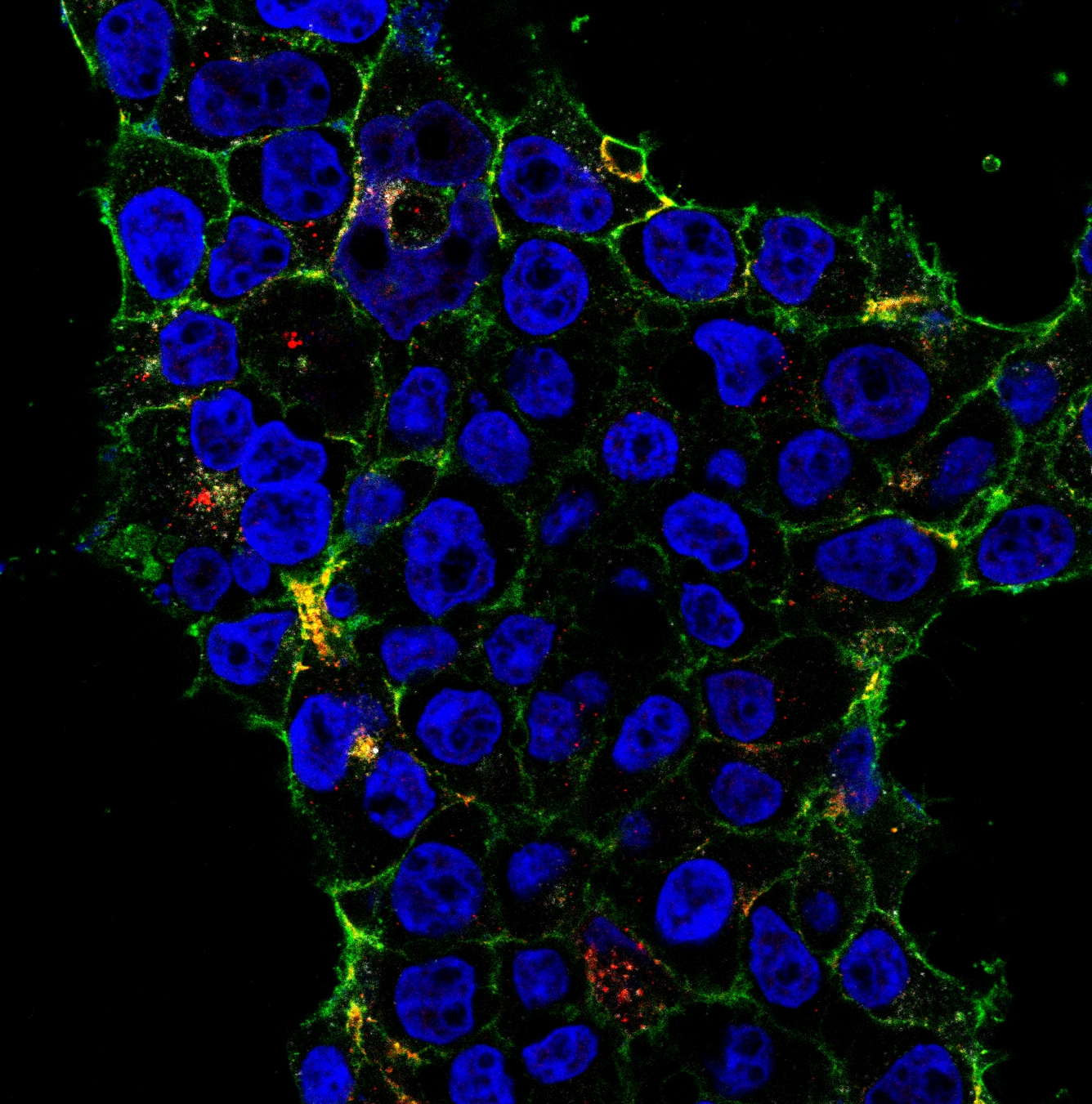
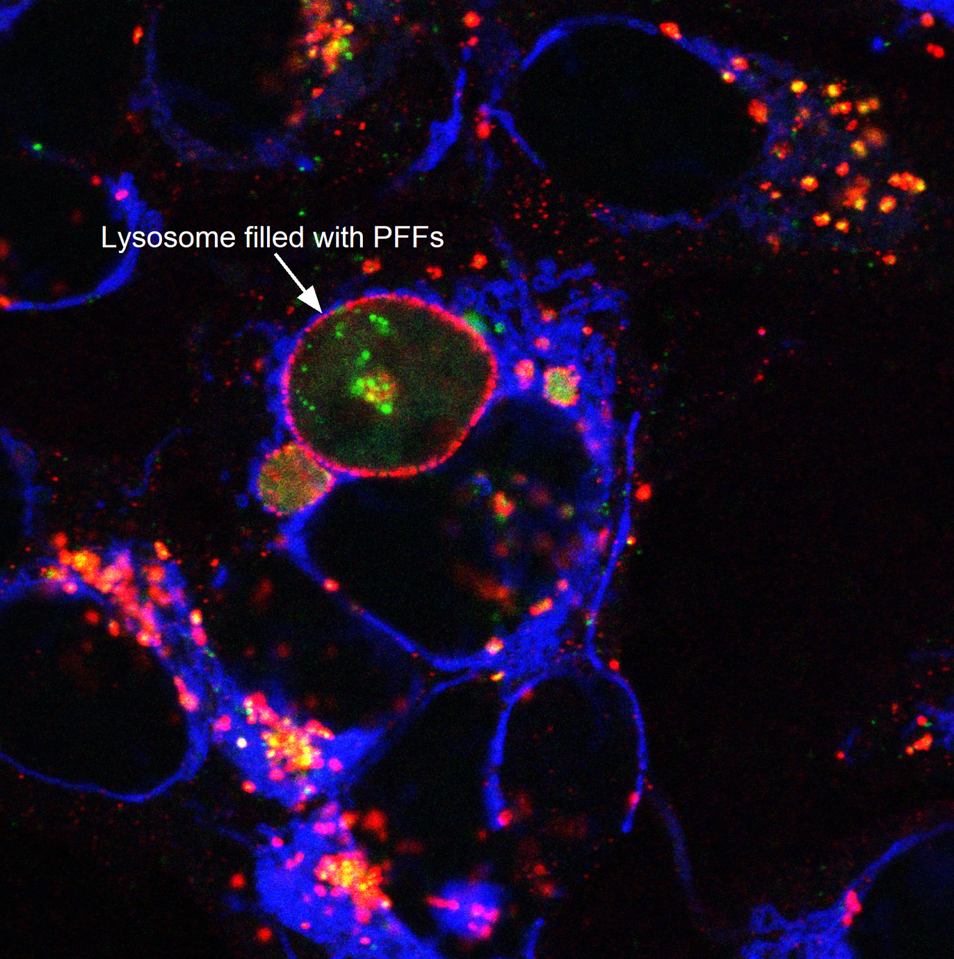
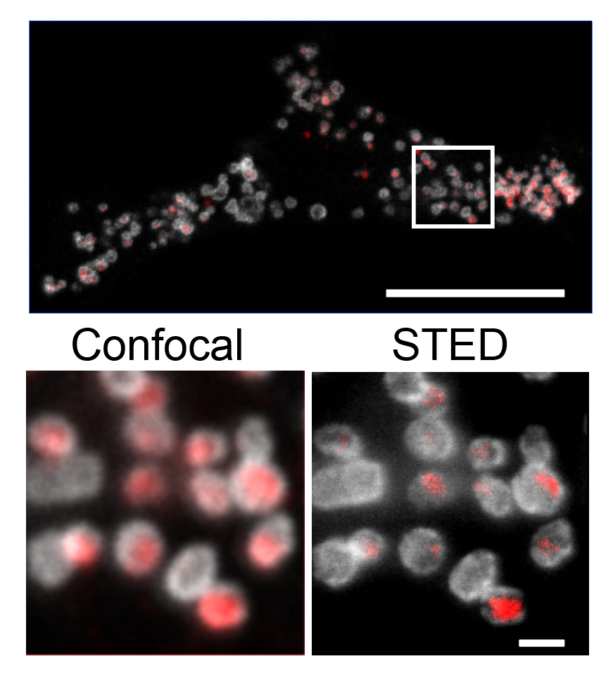
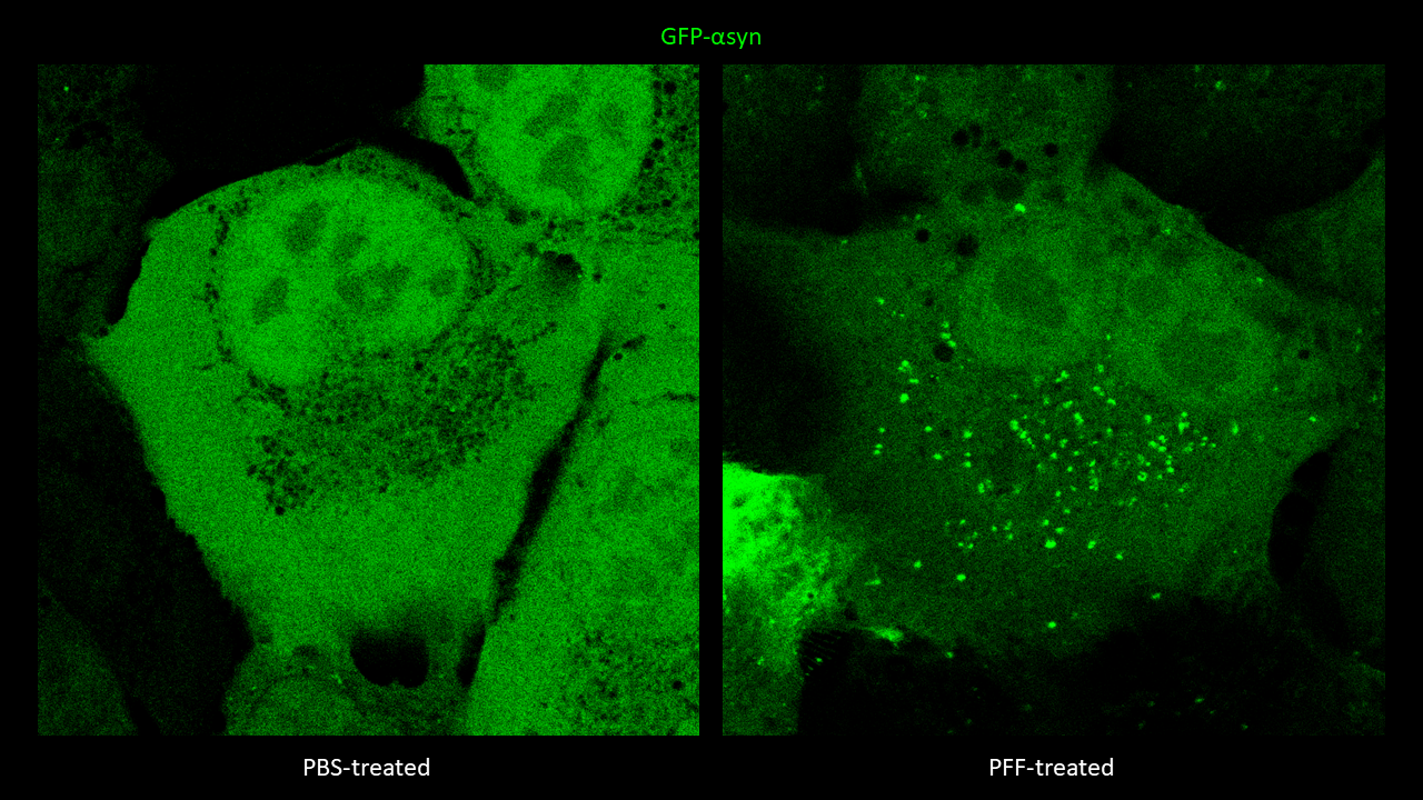
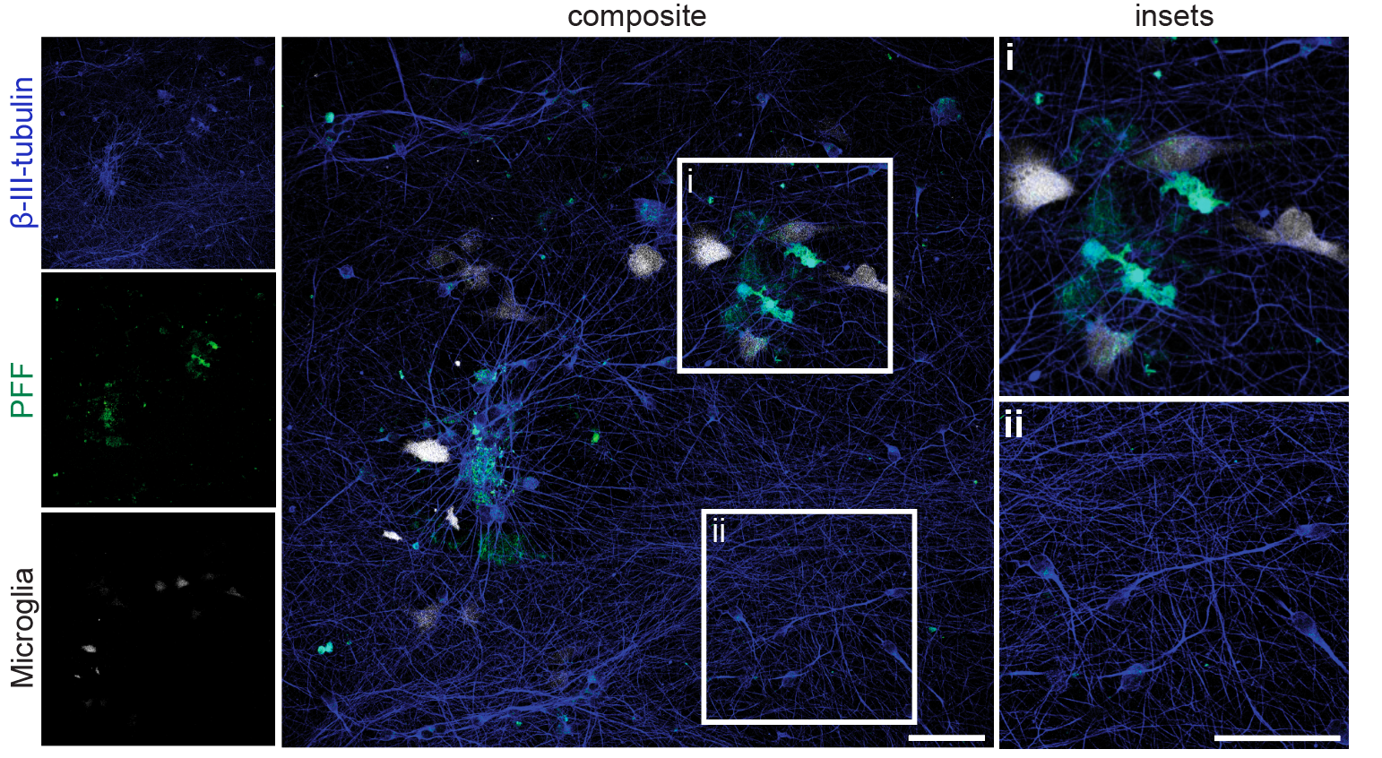
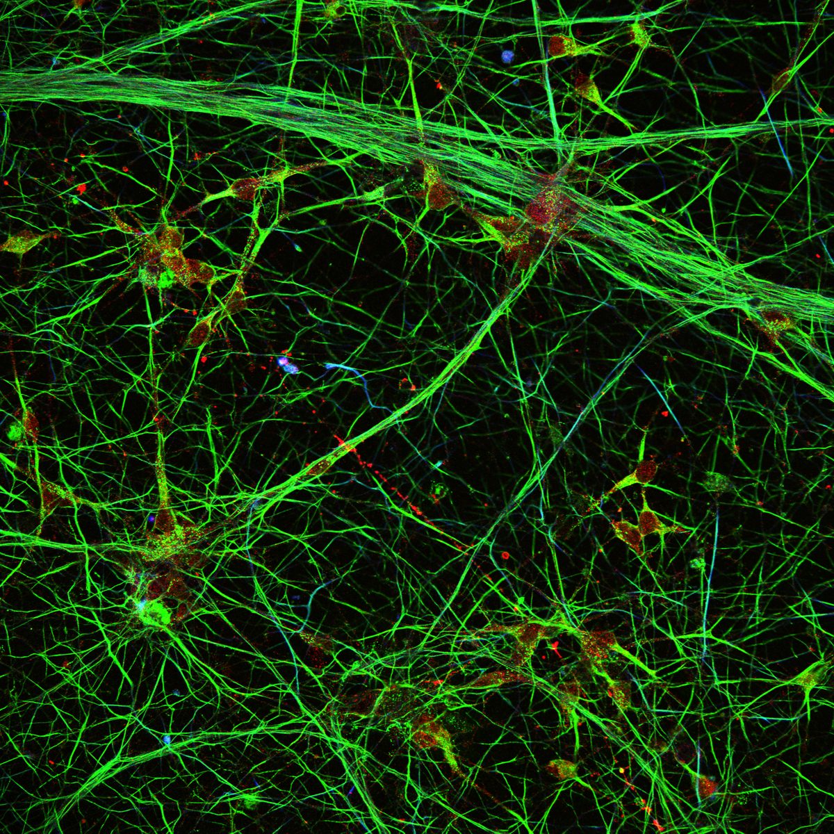
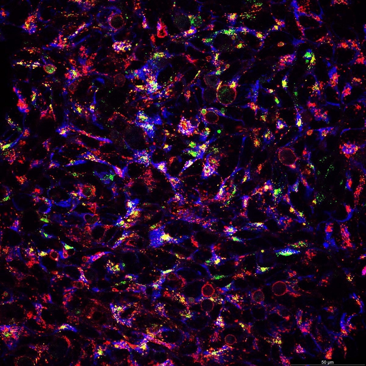
I use advanced microscopy — from live-cell confocal to STED super-resolution, electron microscopy, and high-content platforms — as primary discovery tools to uncover mechanisms in neurodegeneration and infection.
Confocal, STED super-resolution, and transmission EM images in one viewer. Filter by modality, swipe or use the arrow keys, and open any image full screen.
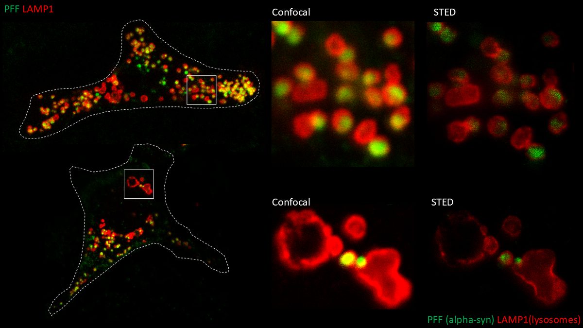
I perform confocal microscopy (Leica) for live and fixed samples, tracking rapid uptake of α-synuclein fibrils and spike protein internalization. STED microscopy (Abberior) resolves sub-lysosomal fibril localization. Zeiss Airyscan provides super-resolution for directional transport and spatial redistribution studies. TIRF (Nikon) for membrane-proximal events. Live-cell imaging captures dynamic processes: fibril internalization, lysosomal swelling, mitochondrial fragmentation, and Parkin translocation.
9 images · Confocal & Super-Resolution GalleryOpen in gallery →
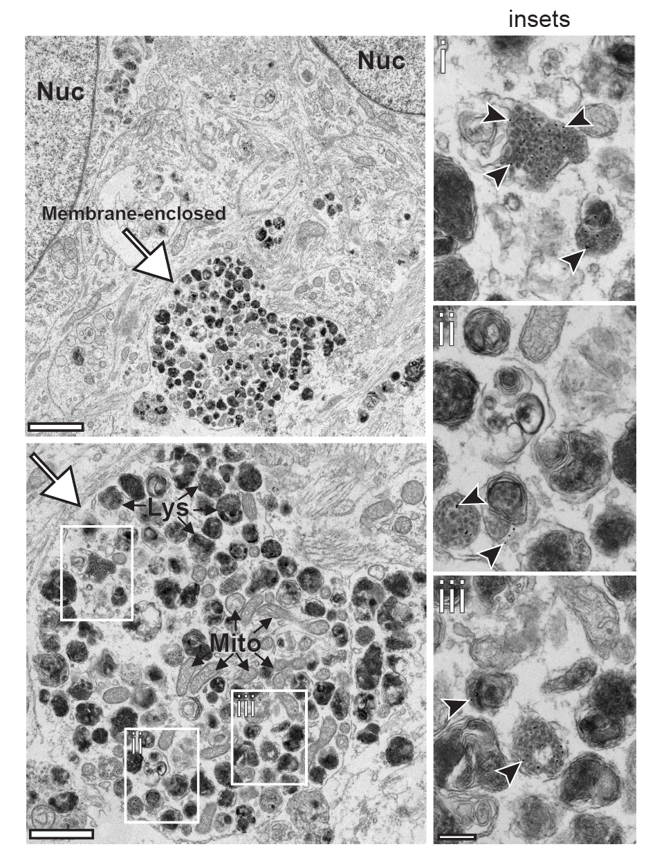
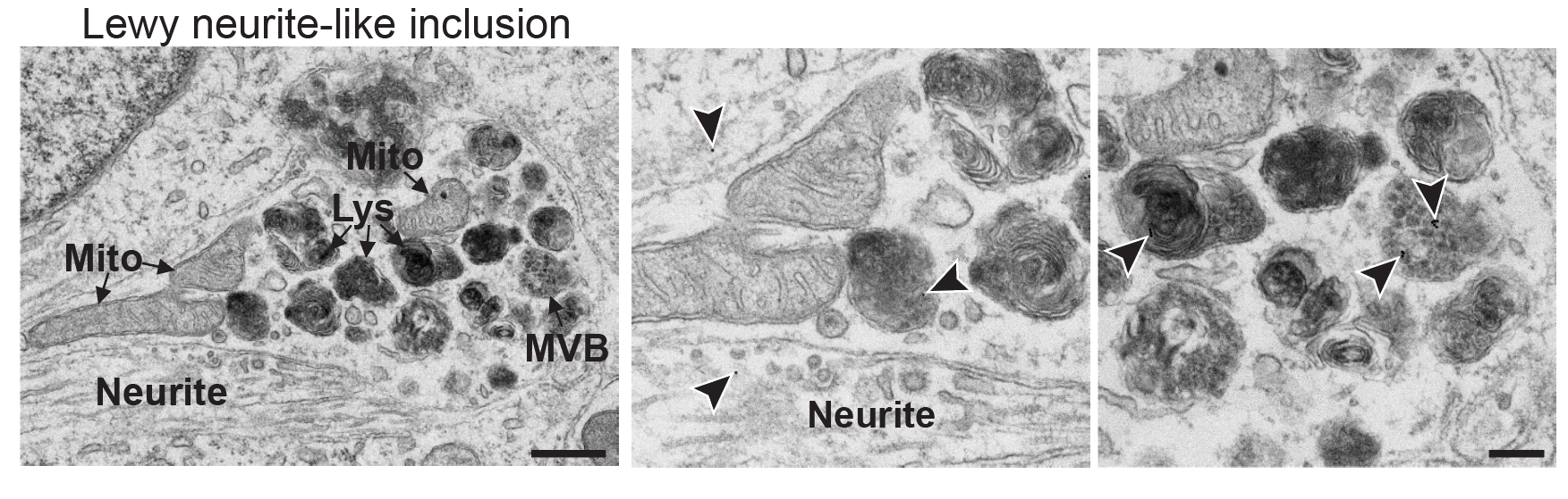
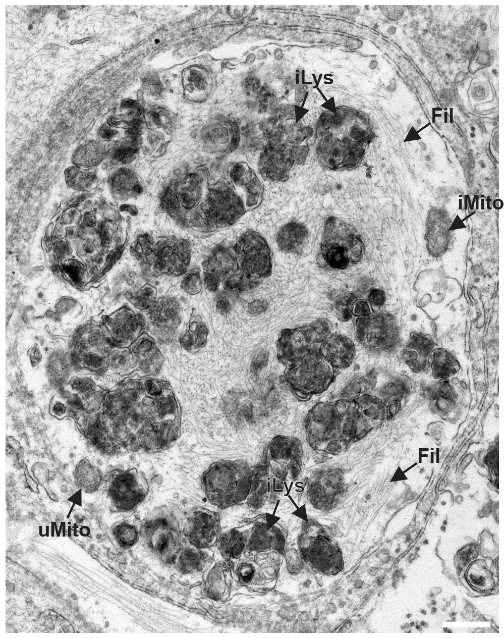
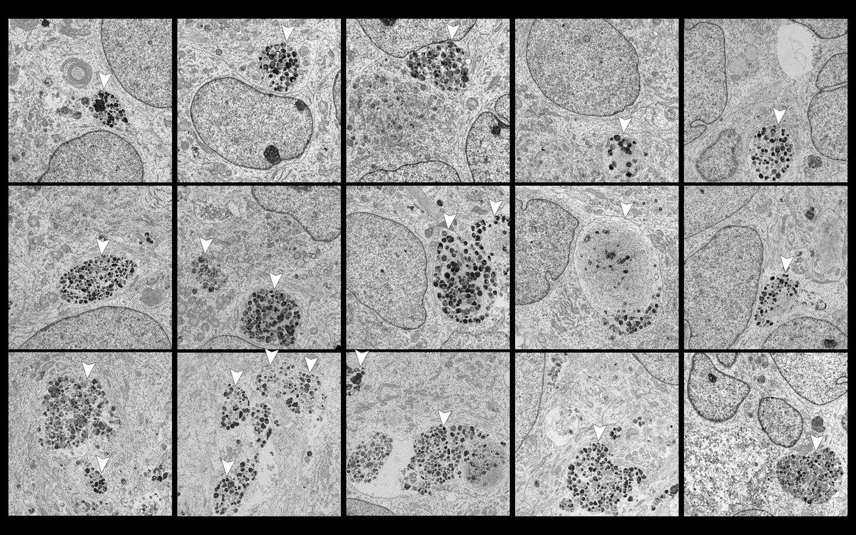
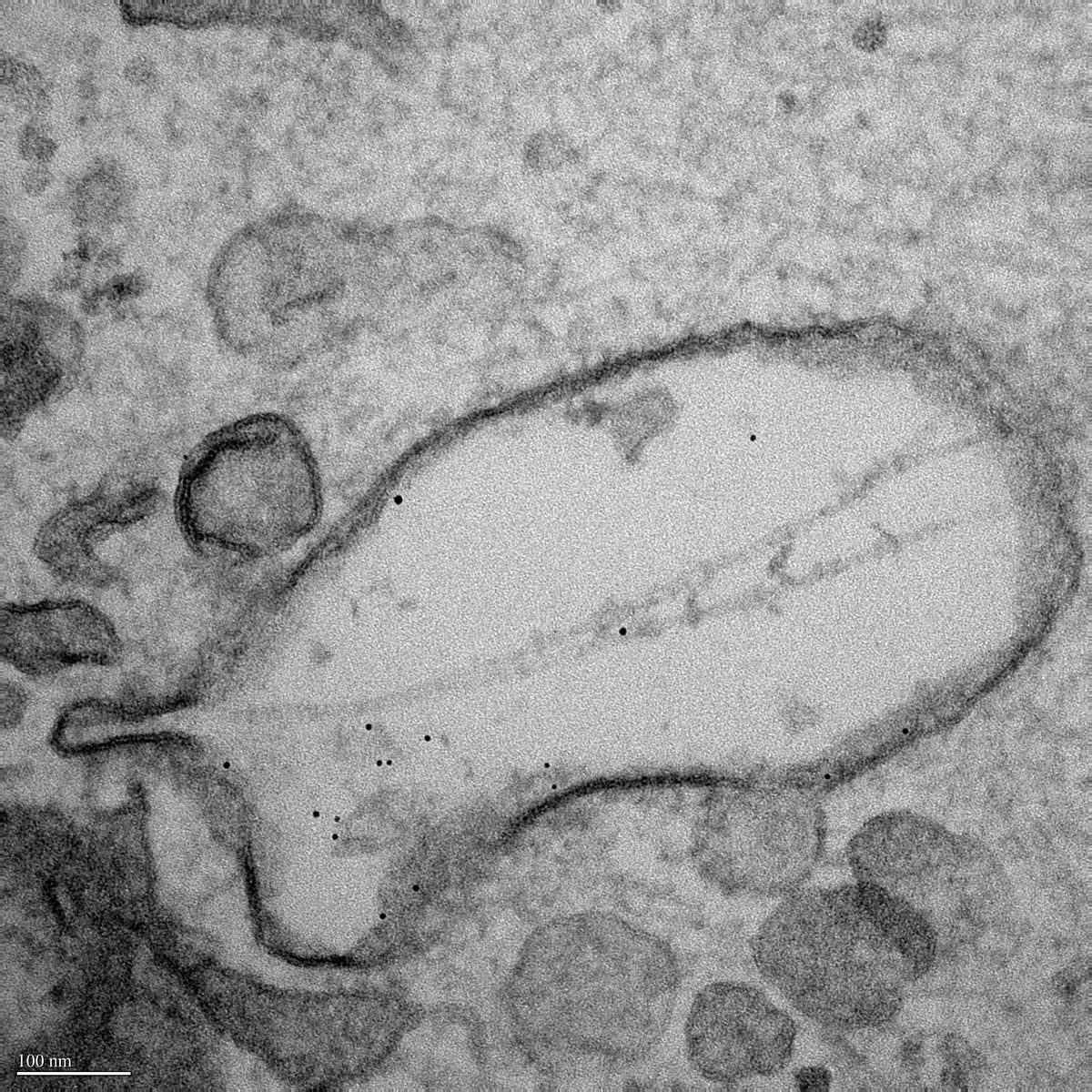
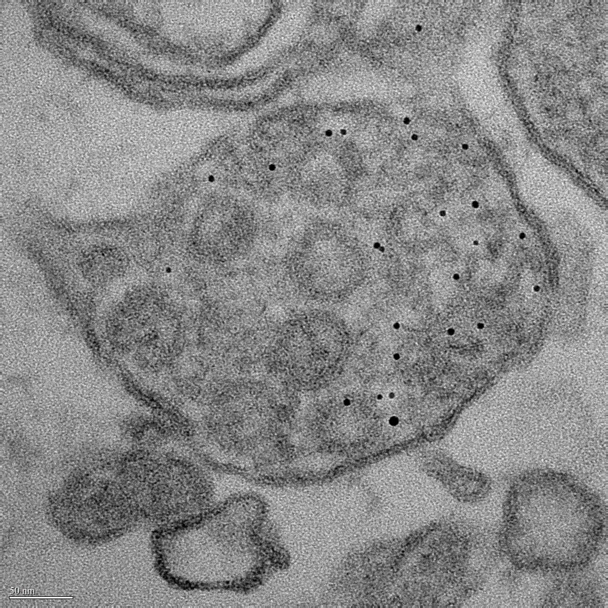
I provided the first ultrastructural evidence of Lewy body–like pathology developing in human neurons in vitro. Inclusions were 5–10 µm, membrane-bound, and perinuclear. Nanogold-labeled fibrils enabled antibody-free visualization of trafficking through macropinosomes, MVBs, and lysosomes. Negative-stain EM for protein aggregation and stability assessment. Correlative light-electron microscopy (CLEM) linking ultrastructure to molecular readouts.
6 images · Electron Microscopy GalleryOpen in gallery →
Immunohistochemistry (IHC) assay development including antibody validation, antigen retrieval optimization, chromogenic (DAB) IHC, and multiplex immunostaining. Quantitative scoring (H-score, percent-positive, intensity thresholds). Tissue handling: cryosectioning, FFPE sectioning, section handling and storage. RNA in situ validation using FISH/RNAscope. Experience with postmortem human tissue and rare sample handling.
I develop and execute high-content imaging assays on Opera Phenix and Incucyte platforms for pharmacological and genetic perturbation screening. Cell painting dyes (LysoTracker, Hoechst, MitoTracker, Cell Mask) enable multiplexed phenotypic readouts across 96- and 384-well formats.
Comprehensive descriptions of key experimental techniques, assay platforms, and analytical methods referenced throughout this page.
I use the Zeiss LSM 880/980 Airyscan detector for ~120 nm lateral resolution imaging (1.7× improvement over standard confocal). Applications include α-synuclein fibril morphology, synaptic vesicle colocalization, and organelle contact site imaging. Airyscan provides the resolution enhancement of structured illumination with the optical sectioning of confocal, making it ideal for thick specimens like iPSC-derived neuronal cultures.
I perform STED (Stimulated Emission Depletion) nanoscopy on the Abberior platform at ~40–60 nm resolution. This enables visualization of protein nanodomains, receptor clustering, and cytoskeletal ultrastructure that are unresolvable by confocal. I have applied STED to image phospho-α-synuclein aggregates within single lysosomes and to resolve individual synaptic active zones in iPSC-derived neurons.
I operate the Nikon TIRF system for single-molecule and near-membrane imaging with ~100 nm axial resolution. TIRF selectively excites fluorophores within the evanescent wave (~100–200 nm from the coverslip), eliminating out-of-focus background. Applications include membrane receptor dynamics, exocytosis/endocytosis events, α-synuclein fibril–membrane interactions, and single-particle tracking of tagged proteins.
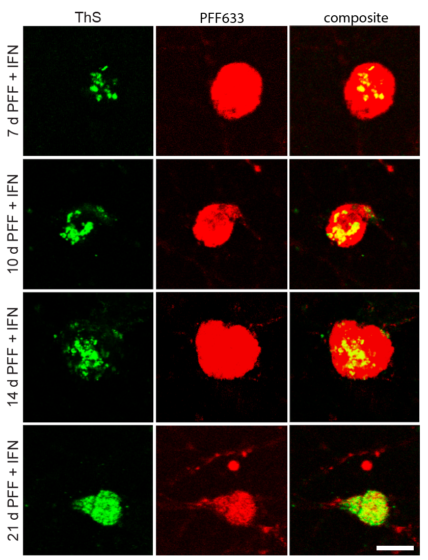
I perform correlative light and electron microscopy (CLEM) to bridge fluorescence imaging with ultrastructural EM. Cells are first imaged by confocal for fluorescent markers (e.g., GFP-LC3, pSyn-488), then processed for transmission EM. Grid-mapped coordinates enable identification of the same cell across modalities. I used CLEM to demonstrate that fluorescent α-synuclein inclusions correspond to membrane-bound, fibril-containing Lewy body–like structures at the EM level.
I operate the Sartorius IncuCyte S3/SX5 for continuous, label-free or fluorescence-based live-cell monitoring inside the incubator. Applications include cell proliferation (phase confluence), cytotoxicity (Cytotox Green/Red), apoptosis (Annexin V-488), neurite outgrowth (NeuroTrack), and immune killing (co-culture real-time lysis). I routinely run 96/384-well plates with 4-hour scan intervals over 7–14 days for longitudinal compound studies.