Image Gallery
Imaging from each part of the neuroimmunology program, collected in one viewer. Filter by topic, swipe or use the arrow keys, and open any image full screen.
Investigating the intersection of innate immunity and neurodegeneration in human iPSC-derived models. My neuroimmunology program dissects how pro-inflammatory cytokines (IFN-γ, TNF-α, IL-1β) drive neuronal vulnerability, α-synuclein aggregation, and lysosomal dysfunction — and how microglia and astrocytes modulate these pathological cascades. This work has yielded first-author publications in Nature Neuroscience and Cell Reports, with direct translational applications for anti-inflammatory neuroprotection strategies.
Imaging from each part of the neuroimmunology program, collected in one viewer. Filter by topic, swipe or use the arrow keys, and open any image full screen.
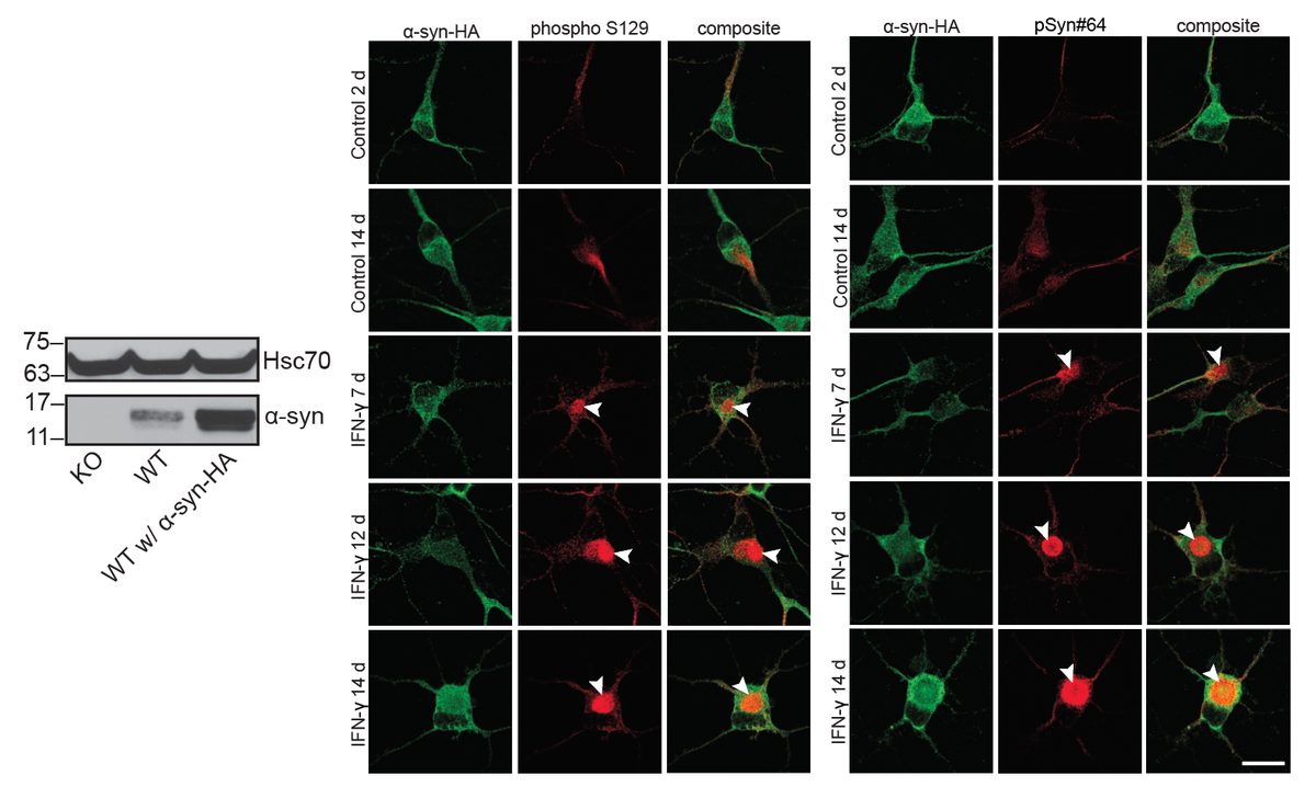
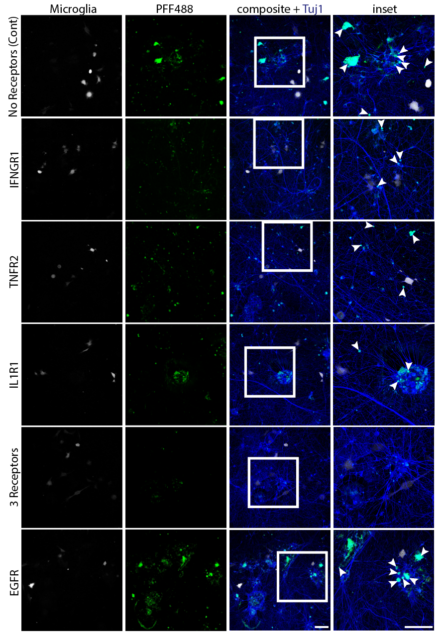
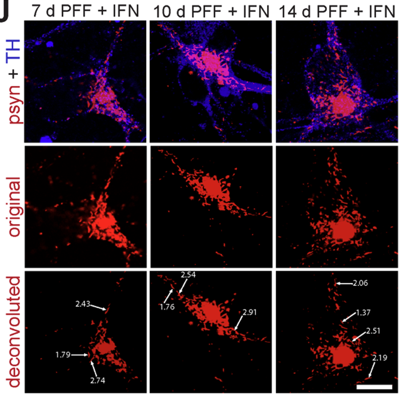
Systematic investigation of how pro-inflammatory cytokines impact iPSC-derived dopaminergic neuron health. IFN-γ exposure induces MHC-I upregulation, antigen presentation machinery activation, and downstream lysosomal dysfunction. TNF-α and IL-1β act synergistically to promote α-synuclein phosphorylation and Lewy body-like inclusion formation. These findings establish a mechanistic framework connecting peripheral inflammation to neuronal pathology in Parkinson's disease.
3 images · Cytokine-Driven Neuronal VulnerabilityOpen in gallery →
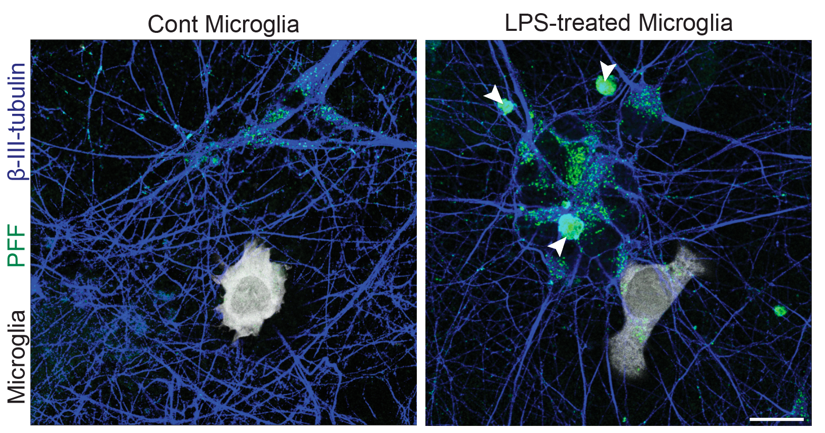
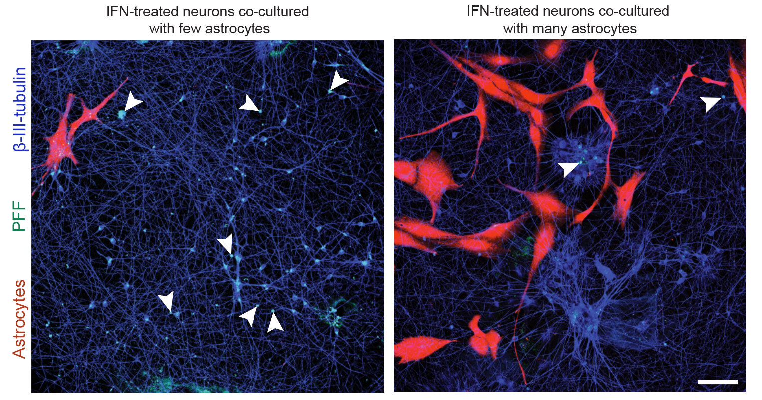
Developed tri-culture systems combining iPSC-derived dopaminergic neurons with microglia and astrocytes to model the neuroimmune microenvironment. Microglia amplify α-synuclein inclusion formation through pro-inflammatory cytokine secretion and direct phagocytic interactions. Astrocytes provide neuroprotection via glutathione transfer and metabolic support, but become neurotoxic when activated by microglial TNF-α. These co-culture platforms enable screening of anti-inflammatory therapeutics in physiologically relevant contexts.
3 images · Neuron–Glia Co-Culture SystemsOpen in gallery →
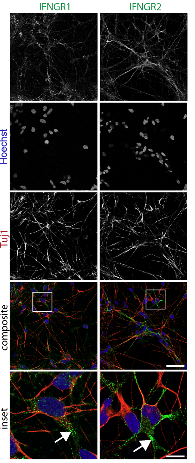
Systematic characterization of cytokine receptor expression and signaling in iPSC-derived dopaminergic neurons. Identified IFNGR1, TNFR2, and IL1R1 as the dominant receptor subtypes mediating inflammatory neurodegeneration. Confocal imaging confirmed surface receptor localization and internalization dynamics upon ligand binding. These receptor maps guided the selection of anti-cytokine biologics (anti-TNF-α, anti-IL-1β, anti-IL-6) that demonstrated dose-dependent neuroprotection.
1 image · Neuroimmune Receptor DissectionOpen in gallery →
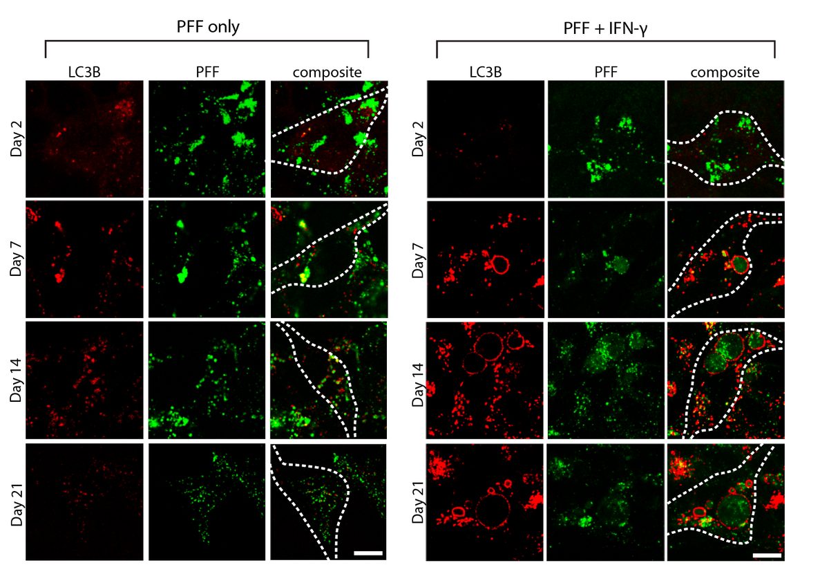
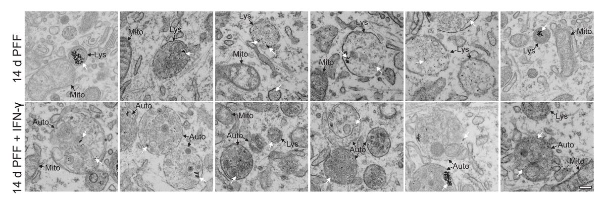
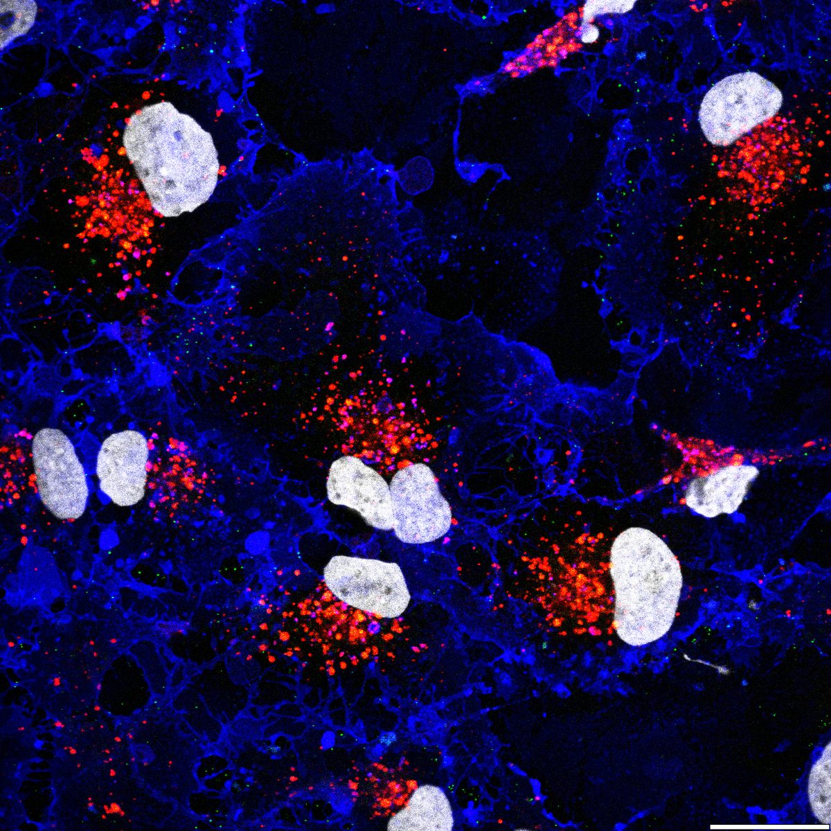
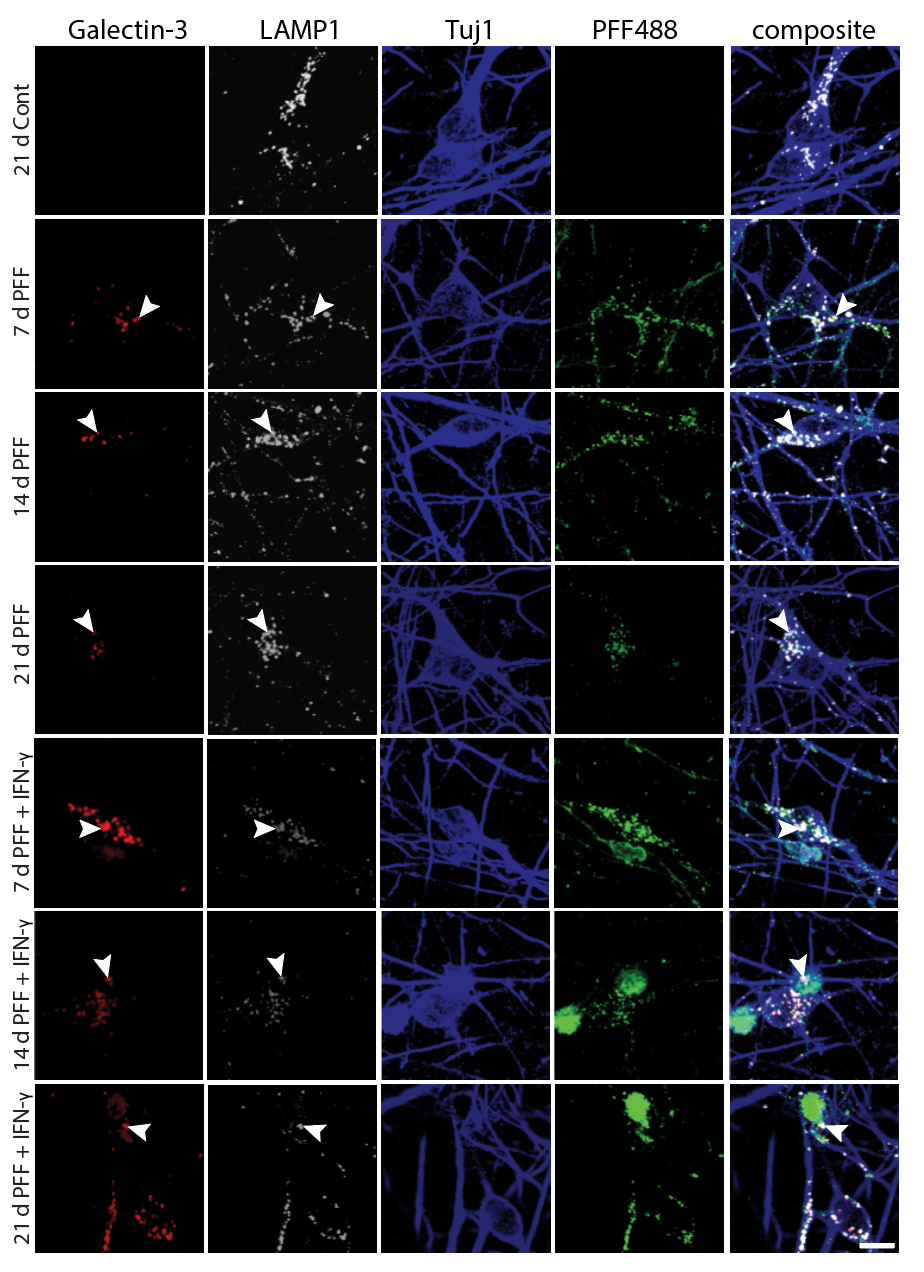
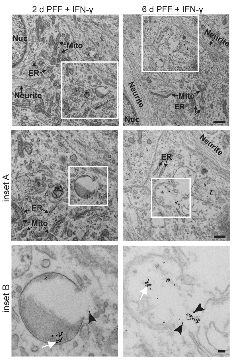
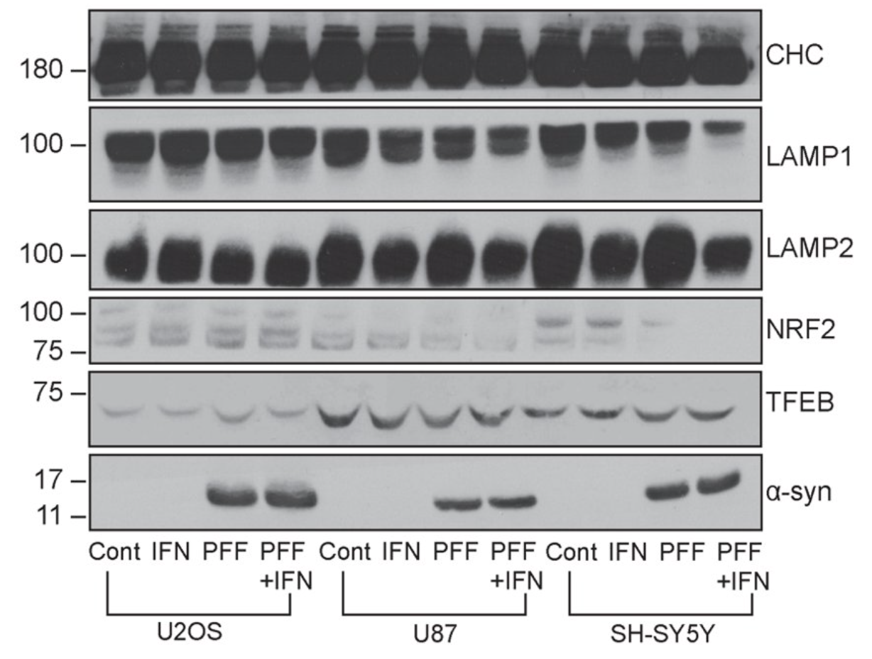
IFN-γ signaling triggers progressive lysosomal dysfunction in dopaminergic neurons, characterized by LAMP1+ compartment enlargement, cathepsin activity reduction, and impaired autophagic flux. This creates a feed-forward loop: dysfunctional lysosomes fail to clear α-synuclein aggregates, which further compromise lysosomal membrane integrity (detected by galectin-3 puncta). Electron microscopy revealed multilamellar body accumulation and lysosomal membrane rupture in IFN-γ–treated neurons.
3 images · Inflammation-Induced Lysosomal DysfunctionOpen in gallery →
Comprehensive MSD multiplex profiling of secreted cytokines, chemokines, and neurodegeneration markers across inflammatory treatment paradigms. Established pharmacodynamic biomarker panels for monitoring anti-inflammatory therapeutic efficacy. Key readouts include phospho-α-synuclein (pS129), NfL, GFAP, and a 15-plex cytokine panel enabling simultaneous quantification of inflammatory and neurotoxic cascades.
2 images · Neuroimmune Biomarker ProfilingOpen in gallery →